



Article Sidebar

Published:
Jul 7, 2026DOI:
10.4314/eajns.v5i3.7Keywords:
Article Details

This work is licensed under a Creative Commons Attribution-NonCommercial-NoDerivatives 4.0 International License.
Main Article Content
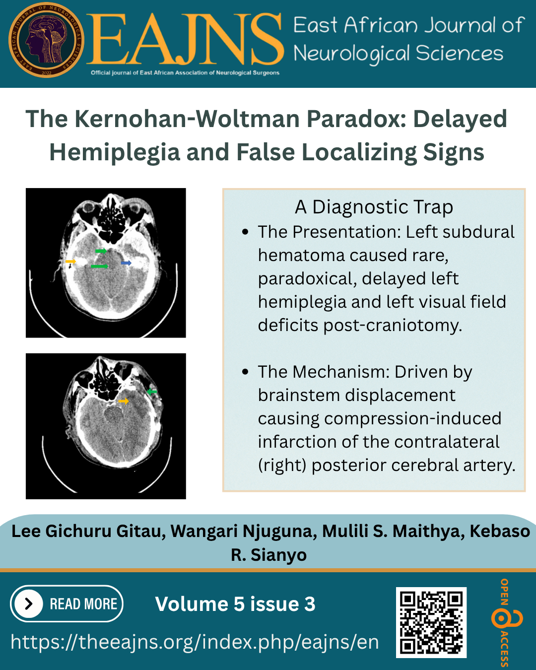
Delayed Hemiplegia and Posterior Cerebral Artery Infarction in Kernohan-Woltman Notch Phenomenon: Case Report and Literature Review
Lee Gichuru Gitau

Wangari Lucy Njuguna
Mulili Solomon Maithya
Kebaso Raymond Sianyo
Abstract
Background: Kernohan-Woltman Notch Phenomenon (KWNP) causes ipsilateral mydriasis and paradoxical ipsilateral hemiparesis or hemiplegia. Delayed ipsilateral hemiplegia and infarction of the posterior cerebral artery (PCA) are rare manifestations of KWNP. Case summary: A 42-year-old man presented with loss of consciousness after an accident. His GCS was 10/15. He was anisocoric with left-sided mydriasis, and had hypertonia and hyperreflexia in the left lower and upper limbs with normal tone and reflexes in the right limbs. A computerized tomography (CT) scan showed an acute left subdural hematoma with midline shift. After craniotomy, he developed hemiplegia (Medical Research Council [MRC] grade 0/5) on his left upper and lower limbs. He also developed an “inability to see” from his left eye. He was discharged 4 weeks later with a GCS of 15/15 and power of MRC grade 5/5 on his right and left upper and lower limbs. Investigations: A pre-operative CT scan revealed an acute left subdural hematoma and midline shift. A post-operative CT scan revealed a contralateral (right) PCA infarct. All laboratory tests conducted were normal. Outcome/Interpretation: The patient had an acute left subdural hematoma with midline shift. The subdural hematoma also caused KWNP with delayed hemiplegia and contralateral (right) PCA infarction, which caused visual changes. Conclusion: KWNP may cause delayed hemiplegia. This presentation may also be accompanied by infarction of the contralateral PCA, causing visual changes. Recognizing this presentation is crucial in diagnostic formulation for hemiplegia and visual changes occurring after decompressive craniotomy. MRI may be useful in diagnosing KWNP and differentiating it from a primary brainstem hemorrhage.